Berkeley Lab researchers have shown that tiny bubbles carrying hyperpolarized xenon gas hold big promise for NMR (nuclear magnetic resonance) and its sister technology, MRI (Magnetic Resonance Imaging), as these xenon carriers can be used to detect the presence and spatial distribution of specific molecules with far greater sensitivity than conventional NMR/MRI. Applications include molecular imaging of complex solid or liquid chemical and environmental samples, as well as biological samples, including the detection and characterization of lung cancer tumors at an earlier stage of development than current detection methodologies.
“Rather than the protons used as the reporting medium in conventional NMR/MRI, our reporting medium is the NMR/MRI-active isotope of xenon (Xe-129),” says chemist Alex Pines, who led this research along with Todd Stevens and Matthew Ramirez.
Pines, a faculty senior scientist in Berkeley Lab’s Materials Sciences Division and the Glenn T. Seaborg Professor of Chemistry at the University of California (UC) Berkeley, is one of the world’s foremost authorities on NMR/MRI.
“Xenon is an ideal reporter because it is inert and nontoxic, and has no background in natural samples,” he says. “It can also be hyperpolarized using established optical techniques, which facilitates detection of xenon NMR contrast agents at sub-picomolar concentrations. This is orders of magnitude below the threshold for detecting proton contrast agents in a conventional NMR/MRI system.”
NMR and MRI are well-established technologies based on an intrinsic quantum property of atomic nuclei called “spin.” While non-invasive and capable of providing information in a wide range of applications, NMR/MRI techniques have long been plagued by a lack of sensitivity. Over several decades Pines has led the development of numerous strategies for hyperpolarizing the spins of atomic nuclei to boost the strength and sensitivity of NMR/MRI signals.
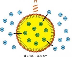
In this latest development, Pines, Stevens and Ramirez combined a strategy called hyperCEST (for hyperpolarized xenon chemical exchange saturation transfer) with nanoemulsions that can be made to bind to molecular targets of interest. These emulsions consist of a perfluorocarbon core and a stabilizing surfactant that form droplets in water measuring between 160 and 310 nanometers in diameter.
“The hyperpolarized xenon is freely exchanged between the aqueous solution and the perfluorocarbon interior of the droplets, which are spectroscopically distinguishable and allow for detection of the xenon signal using a hyperCEST scheme,” Pines says. “With this technique we’re able to detect target molecular concentrations as low as 100 femtomolars in just five seconds.”
The use of hyperpolarized xenon and perfluorocarbon nanoemulsions should prove especially useful for chemical sensing applications in which sparse analytes need to be detected from opaque mixtures that cannot easily be probed with optical-based techniques. Such mixtures include biofuels and other complex fluids, and the surfaces of nano materials and other novel solids. However, from the public’s perspective, perhaps the most intriguing potential application is the early diagnosis of lung cancer.
“Currently, the most widely used technologies for lung cancer diagnostics are X-ray CT and nuclear medicine techniques, all of which use ionizing radiation that precludes frequent screening and longitudinal studies,” says Stevens, who is now with the Western University in Canada.
“Our xenon-based MRI can probe the void space of the lung and the lung tissue directly and via chemical exchange. In addition to earlier detection of cancerous tumors, our technique also has the potential to allow for characterization of the types of cancer cells present. By using a library of targeting molecules on the outside of the nanoemulsion droplets, one could determine what agents are binding and infer information about the cancer cells based on the bound-targeting molecules.”
“The next steps in this effort” Stevens says, “will be to extend control over the size and stability of the nanoemulsion droplets, which will enhance the contrast and distribution of the agents, and to optimize droplet targeting systems for some select applications.”
A paper describing this research has been published in the Journal of the American Chemical Society titled “Nanoemulsion Contrast Agents with Sub-picomolar Sensitivity for Xenon NMR.”
| Attachment | Size |
|---|---|
| 429.26 KB |

